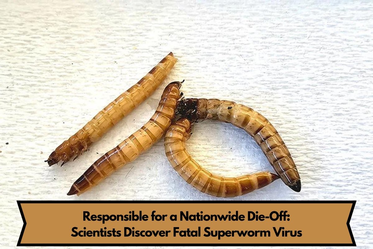
Scientists from Rutgers University-New Brunswick have found a virus that has killed off all superworms across the country. Superworms are a common food source for pets and are also being used more and more as a protein source for people.
A recent article in Cell wrote about this discovery. It gives them a way to look for and spot new viruses and pathogens in plants, animals, and people.
New methods for research in virology
Scientists found what they call the Zophobas morio black wasting virus by using a mix of chopped up beetle bodies and an electron microscope that was kept cool by liquid nitrogen.
The name comes from the fact that the virus is very dangerous to a species of darkling beetle that lives in the subtropics, especially when it just hatches from its eggs and turns into a big brown worm.
Their eggs are about 2 inches long, which makes them bigger than any other species grown as food. That is why they were given the name “superworm.”
How to Solve the Superworm Mystery
A researcher at Rutgers-New Brunswick called Jason Kaelber and a postdoctoral worker at IQB named Judit Penzes worked on the study. Penzes was the first author of the study and Kaelber was an associate research professor there.
“I was trying to find ways to find new viruses that do not depend on DNA or RNA sequencing,” Kaelber explained. “Judit was trying to figure out why beetle farmers were losing all of their superworm colonies to a deadly disease.” “In the end, we found the virus that was killing superworms across the country.”
Penzes, a molecular virologist, was called by beetle farm owners more than a year ago to look into why their superworms were mysteriously dying off at very high rates. Penzes was already well-known in the field for earlier work in which she found a virus that was killing bugs, which are another popular pet food.
How to Do Investigations in the Lab
At first, she bought superworms at pet stores in New Jersey. “My first stop at any pet store was the feeder insect section. I would open the containers and look at the worms,” she said. ”
All of them were sick.” I told the store owners what I saw and said I was studying this virus. I then asked if I could have the tube. They got on board right away. “Take as many as you need,” they told me.
She went back to her lab, got a Magic Bullet mixer, put the worm bodies in it, and blended them very quickly.
The process made a thick mixture of beetle juice, which she then used a way for getting rid of viruses that separates them because of their density. She put a fluorescent light on the spinning tube as the last step. The virus lit up blue.
Also read:-New Research Debunks Violent “Steppe” Invasion Theory in Iberia 4,200 Years Ago
Using Cryo-Electron Microscopy to Find Pathogens
Next, Penzes and Kaelber, another electron microscopist, used a cryo-electron microscope to look at the virus. This type of microscope gives a three-dimensional picture of the virus, including its insides.
Kaelber said, “You freeze a virus, a protein, a cell, etc. so quickly that the water freezes without turning into ice crystals.” “Because we have such high resolution, we can figure out what the amino acid sequence of the protein is without looking at the DNA.”
They used the Protein Data Bank database at Rutgers to match the protein’s structure to all known proteins. They found that it is similar to a virus that affects cockroaches but not exactly the same. It is in the same family as parvoviruses, which are viruses that infect animals.
Concluding Remarks on Virus Research Impact
Researchers found a way to keep the Z. morio beetles from getting sick by treating them with a closely related virus from a different species that does not make them sick. Based on that work, they are making a vaccine.


